How Heart Works
- Details
- Last Updated: Sunday, 24 November 2024 17:54
- Published: Wednesday, 24 May 2023 21:53
- Hits: 1044
Heart:
Heart is one of the central system keeping the human body alive. The instant it stops working, you go dead. Heart failure is the number one cause of death.
Here's a good link to Heart's working: https://www.youtube.com/watch?v=_qmNCJxpsr0
One more good one with hand drawn diagram by Dr John Campbell: https://www.youtube.com/watch?v=VWamhZ8vTL4
Heart explained with a physical 3D model: https://www.youtube.com/watch?v=WyKBFWYKRBY
There's artificial hearts being made, which are someday going to replace failed hearts all together. The longest such an artificial heart has worked continuously in a human body is about 4 years. So, there's hope that over the next few decades, we may have artificial hearts lasting 20-30 years. Here's a video on such artificial hearts: https://www.youtube.com/watch?v=3uV8XZcIBbk
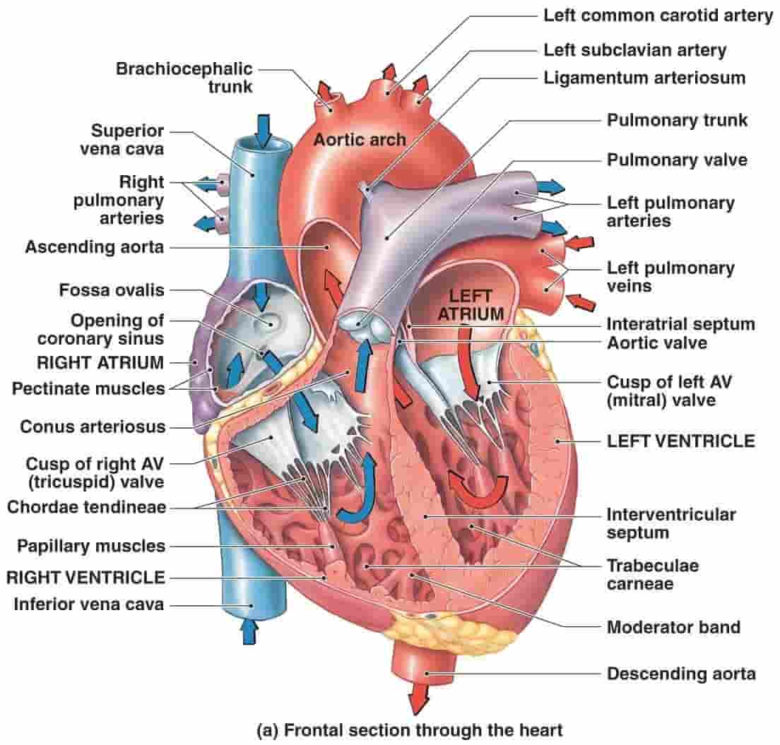
Heart anatomy: Heart sits in the center of our body near chest region. However, we hear that Heart is to our left. This is because some bigger chamber of heart is little bit to the left, so looks like heat is to the left. Right half of Heart is the one that collects dirty deoxygenated blood and passes it to the lungs for oxygenation. Once it gets oxygenated by lung, it's pushed to the left side of heart which transfers it to the rest of the body.
Link => https://medicoapps.org/wp-content/uploads/2018/10/1522314495.jpg
4 chambers of heart:
- Right Atrium (RA): Deoxygenated blood from all over the body is passed via blood pipes called Vena Cava (Superior Vena Cava (SVC) for top part and Anterior Vena Cava (AVC) for bottom part). These pipes or Veins enters the RA chamber here. Blood is passed to Right Ventricle which is sitting below the right atrium. There is a valve called "Tricuspid valve (TV)" that opens or closes to allow blood to pass from right atrium to right ventricle.
- Right Ventricle (RV): Deoxygenated blood flows from Right atrium to right ventricle and is pumped out to Lungs for oxygenation. There is a valve called "Pulmonic valve (PV)" that opens or closes to allow blood to pass from right ventricle to the lungs. The pipes going out of the heart to the lungs are called Pulmonary Artery (PA).
- Left Atrium (LA): Oxygenated blood from lungs is passed via blood vessels called Pulmonary Veins (PV). These pipes or veins enter the LA chamber here. The blood is then pushed to Left Ventricle which is sitting below the left atrium. There is a valve called "Bicuspid valve (BV)" or "mitral valve (MV)" that opens or closes to allow blood to pass from left atrium to left ventricle. MV is the equiv of TV that's seen on right side of heart b/w RA and RV.
- Left Ventricle (LV): Oxygenated blood flows from left atrium to left ventricle and is pumped out to the whole body (from brain to legs). Blood gets to cells which take oxygen from the blood, which gets the blood deoxygenated. Then this deoxygenated blood is passed on to the right atrium, and the 4 step cycle repeats again. There is a valve called "Aorotic valve (AV)" that opens or closes to allow blood to pass from left ventricle to the Aorta (a thick blood pipe which are arteries) which eventually branches out to the whole body. AV is the equiv of PV that's seen on right side of heart b/w RV and PA.
Atrium are much smaller than Ventricle, so when you see a pic of heart, you mostly see 2 front ventricles. The atriums are on the back and on the top of the heart (Ventricles are on the bottom). Though the cycle repeats from step 1 to step 4, processes in right side of heart are going in parallel to left side of heart, implying right and left atrium beat at the same time, while right and left ventricle also beat exactly at same time. So, we can consider only right side of heart for understanding purposes.
Veins and Arteries:
It's important to understand the difference b/w the two. Both are referred to as the blood vessels in the body. Veins are blood vessels going to the heart, while arteries are blood vessels going out of the heart. Veins get into the heart from 2 places => one from SVC/AVC into RA, and other from the lungs to LA in heart. Arteries get out of the heart from 2 places => one from RV going to the lungs, and other from the LV in heart. Usually blood vessels going into the heart carry deoxygenated blood, so veins are shown blue in color (blue means deprived blood, i.e in Hindi, people say a person's body has turned blue or "neela"meaning blood supply is dying), while blood vessels going out of the heart carry oxygenated blood, so arteries are shown red in color (red means rich blood). The only exception to this is the blood vessels going to the lungs and coming out of the lungs. The PA goes out of the heart to lungs carrying deoxygenated blood, and is shown blue even though it's an artery. Similarly, the PV goes from lungs to the heart carrying oxygenated blood, and is shown red even though it's a vein.
Blood Flow:
- Right Heart: SVC/AVC (Veins) --> Heart (RA) -> Valve (TV) --> Heart (RV) -> Valve (PV) --> PA (Artery)
- Lungs: --> Lungs -->
- Left Heart: PV (Vein) -> Heart (LA) -> Valve (MV) --> Heart (LV) -> Valve (AV) --> Aorta (Artery)
Arteries: Arteries carry blood away from the heart to the rest of the body. It needs pressure to push blood to everywhere in body. So, they have thick walls with muscle tissue. That thick pipe carrying blood from the heart to body is called "Aorta" as it's a Artery. It's the widest tube you find anywhere in body. It goes until the neck (which is a very short distance), before it branches off into smaller arteries to go to the head. The Aorta then curves back down to your chest. It continues through your abdomen and ends at your groin. Along the way, it splits off into other arteries that deliver oxygen-rich blood to your arms, legs, and the rest of your body..
Veins: Veins push deoxygenated blood back to your heart. Veins have thinner walls and are not as wide as arteries. Unlike arteries, veins generally need to work against gravity to push blood back to your heart. Veins have valves to help with this. These are one-way pairs of flaps inside a vein. They open for blood that’s heading upwards toward the heart, and close to keep blood from flowing back downwards. Your veins usually hold about 75% of all the blood flowing through your body. Your largest veins are the superior and inferior vena cava. Your superior vena cava carries blood from your upper body to the heart. Your inferior vena cava carries blood from everywhere below your heart. Like arteries, these two veins branch off into many other veins throughout your body. The veins coming out of right hear to go to Lungs are called Left Pulmonary Vein and Right Pulmonary vein. Muscle surrounds most veins in your body. When you walk, run, or otherwise use your muscles, they make a squeezing motion. These squeezes push against the vein and force the blood upwards toward your heart.
Capillaries: Arteries and veins connect through structures called capillaries. Capillaries are small webs of thin tubes that connect to an artery on one side and a vein on the other. Some parts of your body have more capillaries depending on how much energy they need. For example, your muscles use a lot more energy than your skin, which is why your muscles have more capillaries than your outer skin.
Heart Disease:
CVD (Cardiovascular disease) refers to all diseases of cardiovascular system – which consists of the heart and all the blood vessels in the body. CVD is leading cause of death globally, with 18M deaths every year.
- Atherosclerosis: It is the main underlying cause of CVD. It is a disease in which plaques consisting of fat, cholesterol, calcium and other substances build up in the walls of arteries. Over time, the plaques harden, narrowing the opening of the arteries and restricting blood flow. If atherosclerosis occurs in one of the arteries that supply blood to the heart, it can cause a Heart Attack. If blood clot (known as thrombosis) occurs in one of the arteries to the brain, it causes a Stroke. Heart Attack and Cardian Arrest are used interchangeably, though they are different. A cardiac arrest usually occurs when there is a malfunction in the heart’s electrical system that causes the heart to stop beating properly. It suddenly stops beating. This results in the stoppage of blood flow to the brain and other vital organs, and the sufferer loses consciousness and stops breathing normally, resulting in death.
- Stroke (Aka Brain Stroke or Brain attack): 16M ppl suffer from stroke every year. Just a few hours of poor blood flow to some parts of brain causes the body to become paralyzed forever. Few symptoms of stroke include face changes, unbalanced walking, difficulty speaking or slurred speech, etc. 2 kinds of stroke:
- Ischemic Stroke: This is caused due to lack of blood flow (because of a clot in the blood artery to brain). It can be temporary called as Transient Ischemic Attack (TIA) or permanent called as Ischemic Stroke. There is no medication for this, although some medicines given within few hours of stroke have been said to help dissolve the clot. However, there are medicines that are actively being researched to give to survivors of a Brain stroke so that they they form any more blood clots in future. One such medicine => https://www.hri.org.au/news/groundbreaking-world-first-trial-offers-new-hope-to-stroke-sufferers
- Hemorrhagic stroke: This is caused when a blood vessel in your brain ruptures or breaks, spilling blood into the surrounding tissues. This is very deadly and untreatable. However this is less common.
- Stroke (Aka Brain Stroke or Brain attack): 16M ppl suffer from stroke every year. Just a few hours of poor blood flow to some parts of brain causes the body to become paralyzed forever. Few symptoms of stroke include face changes, unbalanced walking, difficulty speaking or slurred speech, etc. 2 kinds of stroke:
- Arrythmia (Irregular heartbeat): t is a type of heart condition where the heart rhythm is not steady. Instead, the heart may feel as though it were skipping a beat, have extra heartbeats every now and then, flutter or race, or beat faster or slower than normal. There are many types of arrhythmias, varying in severity and danger. Atrial Fibrillation (AF) is the most common type. Problems with the heart’s rhythm can occur when there is any interruption to the electrical signals that stimulate the heart’s pumping activity.
- Congestive heart failure (CHF): CHF is #1 heart disease. It is a broad term used to describe a clinical condition resulting from the inability of the heart to adequately pump blood and causing symptoms such as orthopnea, dyspnea on exertion and edema.Heart has blood filling in and blood pumped out every beat. Insufficient filling or insufficient pumping are both problems that lead to CHF.
- Diastolic dysfunction, which is a condition of impaired ventricular filling. Less common.
- Systolic dysfunction, which is a condition of impaired ventricular emptying.
- Ejection Fraction (EF) : of heart is defined as volume of blood pumped per beat out of heart's chamber to the volume of blood filled per beat into the heart's chamber. This is defined for both left ventricle (LVEF) and right ventricle (RVEF). The full capacity of heart is about 120ml-150ml for an adult, and after each beat, volume of blood remaining is ~50ml, meaning 70ml-100ml was pumped out per beat giving an EF=60%-65%. EF of 50%-70% is considered normal. Low LVEF of < 40% is the most common kind of CHF (known as HFrEF) as it results in insufficient supply of blood to body parts. Low RVEF means heart is not pumping enough blood in lungs resulting in various issues. Usually, HF refers to tlow LVEF.
Treatment plan for HFrEF (Heart Failure with reduced Ejection Fraction):
These are 4 pillars of this treatment, which cosnsists of 4 different medications introduced in parallel.
4 Pillars:
- Beta Blocker: This is the first pillar. These medications have been around for 30+ years.These are first line of defense.
- There are several types of beta-blockers, but only three are approved by the FDA to treat heart failure:
- Bisoprolol (Zebeta) => Bisoprolol (Zebeta) is a selective beta1 antagonist without significant intrinsic sympathetic activity or vasodilating properties
- Carvedilol (Coreg) => Carvedilol is a non-selective beta-blocker with additional alpha1-blocking and antioxidant activities. Nebivolol is a novel beta-blocker with both a greater degree of selectivity for beta-1 adrenergic receptors than other agents in this class and an ability to stimulate endothelial nitric oxide production, leading to vasodilation and other potential clinical effects. Carvedilol (Coreg) is a novel agent with antagonist activity against alpha1, beta1 and beta2 receptors, as well as some antioxidant activity. It is the only beta blocker labeled by the U.S. Food and Drug Administration (FDA) for the treatment of heart failure.
- Metoprolol (Toprol) => Like bisoprolol, metoprolol tartrate (Lopressor) and metoprolol succinate (Toprol XL) are beta1-selective blockers without significant intrinsic sympathetic activity or vasodilating properties.
- A study published in 2000, showed little benefits for all 3 drugs when measured in terms of mortality rates (only 40% fewer deaths, but => https://www.aafp.org/pubs/afp/issues/2000/1201/p2453.html
- There are several types of beta-blockers, but only three are approved by the FDA to treat heart failure:
- ARNI inhibitor: ARNI is a combination of sacubitril, a neprilysin inhibitor, and valsartan, an angiotensin II receptor blocker (ARB) and is now considered first choice for initiation. ex: Vyamada (sold in India).
- SGLT2 inhibitor: This has an additive when added on top of ARNI inhibitor. Two drugs, dapagliflozin or empagliflozin are recommended to reduce the risk of heart failure hospitalization and cardiovascular death in HFrEF patients.
- MRA: Aldosterone Receptor Antagonist are also known as MRA. Discovered in the 1990s, these drugs are aldosterone antagonists that reduce excess fluid in the body while preventing the loss of potassium. Yet the primary efficacy is from neurohormonal blockage. They are weak diuretics. The normal dose is usually 25 mg daily.